-
FALLOT'S TETRALOGY
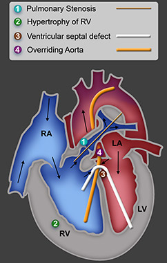
SPECIMEN II:i:8 SD508/1940 This specimen shows the four components of the tetralogy of Fallot: 1.Sub-pulmonary stenosis (stenosis just below the pulmonary valve), indicated by the thin orange probe. 2.Hypertrophy of the right ventricle, a consequence of the obstruction to the pulmonary outflow. 3.Ventricular septal defect; the bent white probe passes from the left to right ventricle through this defect. 4.Overriding aorta (the aorta lies over the ventricular septal d efect and connects to both left and right ventricles),shown by the large orange probe which passes from the right ventricle into the aorta. More info
-
Probe runs from the right ventricle into the aorta, demonstrating the "overriding aorta".
-
The wall of the right ventricle is markedly hypertrophic and thicker than that of the left.
-
The bent white probe passes from the left to right ventricle through the ventricular septal defect.
-
This probe runs from the right ventricle through the stenotic pulmonary valve into the pulmonary trunk.